Scanning Electron Microscope How Does It Work
Ever wonder what a dust mite's face really looks like? I mean, beyond the blurry photos that make you want to burn your mattress?
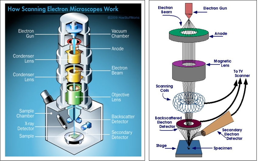
Enter the Scanning Electron Microscope (SEM). It's basically a super-powered magnifying glass on steroids. But instead of light, it uses electrons. And that's where the fun begins.
Shooting Electrons: It's More Fun Than It Sounds
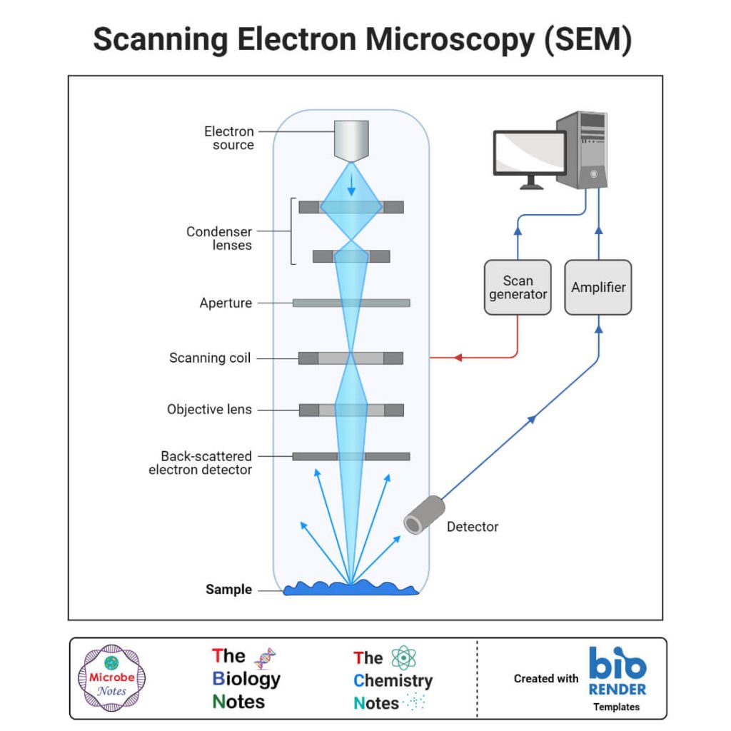
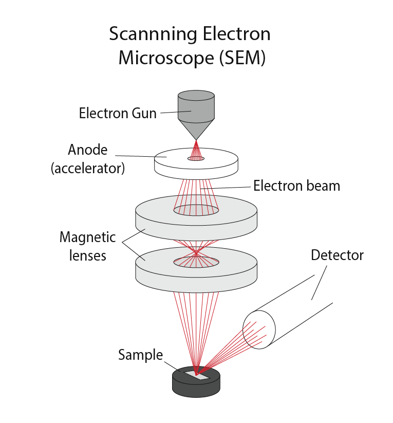
Imagine a tiny, super-focused beam of electrons. Think laser pointer, but instead of light, it's a stream of zippy, negatively charged particles.
Must Read
This beam scans back and forth across whatever you want to see up close. Like a tiny electron lawnmower, but instead of grass, it's exploring the microscopic topography of a Cheeto puff.
Unpopular opinion: "Scanning" sounds way cooler than it actually is. It's not like they're checking your luggage at the airport. More like painting with electrons.
Coating Your Sample: Because Electrons Don't Like to Be Alone
Now, most things don't conduct electricity very well. Think of trying to run electricity through a rubber chicken.
So, before you blast your specimen with electrons, you usually need to give it a thin coat of metal. Gold is a popular choice.
It's like giving your sample a tiny, shimmering suit of armor. Fancy, right?
Seriously though, this coating lets the electrons flow freely and prevents "charging" artifacts. Trust me, you don't want your sample to build up static electricity at that scale. Things get weird.
Detecting the Echoes: Listening to the Electron Chatter
When the electron beam hits your sample, it doesn't just disappear. It's like throwing a bouncy ball at a wall.

Some electrons bounce back (backscattered electrons). Others knock loose other electrons from the sample (secondary electrons).
These ejected electrons are like tiny messengers. They carry information about the surface features of your sample. It's like listening to the echoes to figure out the shape of a cave.
Special detectors around the sample pick up these electrons. They measure the number and energy of the electrons.
This data is then sent to a computer, which builds an image.
Building the Image: From Electrons to Art
The computer uses the information from the detectors to create a grayscale image. Think of it like painting by numbers, but with electrons and a very detailed key.
Areas that reflect more electrons appear brighter. Areas that reflect fewer electrons appear darker.
And voila! You have a super-magnified image of your sample.
They are always grayscale, but scientists can add color later using image processing software. It's like giving a black and white movie a modern makeover.
Unpopular opinion: I actually prefer the grayscale images. They feel more...scientific. More authentic. Like I'm really seeing the raw data.
Magnification: Zooming in on the Unseen
SEMs can magnify things a lot. We're talking tens of thousands, even hundreds of thousands of times.
You can see things that are way too small to see with a regular light microscope. The details are incredible.
Suddenly, the surface of a butterfly wing looks like a landscape from another planet. A grain of sand becomes a mountain range.
It's a bit mind-blowing, honestly.
Why Bother? The SEM's Superpowers
So, why do we need these fancy electron microscopes? What's the point of looking at things so small?
Turns out, there are tons of reasons. They're used in all sorts of fields.
From materials science to biology, SEMs help us understand the world around us at a fundamental level.
Materials Science: Inspecting the Building Blocks
Materials scientists use SEMs to study the structure of metals, ceramics, and polymers. They are looking for cracks, defects, and other imperfections.
This helps them to develop stronger, more durable materials. Like bulletproof vests made from spider silk (okay, maybe not yet).
They can examine how materials behave under different conditions. It helps them to understand how a bridge will hold up in an earthquake, or how a new type of plastic will react to sunlight.
Biology: Peeking Inside Cells
Biologists use SEMs to study the structure of cells, tissues, and organs. They look at the surfaces of cells, to see how they interact with each other.
This helps them to understand how diseases develop. They are also developing new treatments.
They can also identify different types of cells. It's like having a microscopic fingerprint scanner.

Forensic Science: Solving Crimes with Electrons
Forensic scientists use SEMs to analyze evidence from crime scenes. They examine fibers, hairs, and other tiny particles.
It helps them to identify suspects. They reconstruct events. It's like being a microscopic detective.
They can even determine the type of soil found on a suspect's shoes. That's how they connect them to a specific location.
The Future of SEM: Smaller, Faster, Sharper
The technology behind SEMs is constantly improving. Scientists are always developing new and better ways to image things at the nanoscale.
New types of detectors are being developed. They are more sensitive. The new types of detectors can capture more information.
This will allow us to see even smaller details and learn even more about the world around us. Maybe we'll finally get a truly clear picture of that dust mite.
Unpopular opinion: I think the coolest advancement would be a portable SEM. Imagine whipping one out at a party to analyze the molecular structure of the cheese and crackers. Now that's a party trick!
So, next time you see a stunning image taken with a SEM, remember the journey those electrons took. They bounced off a sample. They were captured by a detector. They were turned into a work of art. It's a pretty amazing process, wouldn't you agree?